Thursday, August 27, 2009

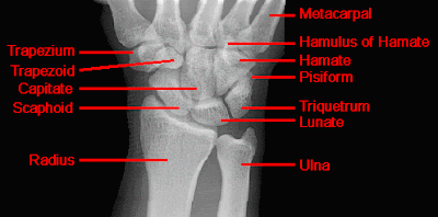
WRIST - NORMAL ANATIOMY

 
FIGURE 2 
 
FIGURE-3 
FIGURE-4 
FIGURE-5

Thursday, August 20, 2009

NORMAL PELVIC ANATOMY


AP VIEW OF NORMAL PELVIS


AP VIEW OF PELVIS SHOWING IMPORTANT ANATOMICAL LINES
  • The five bones that comprise the pelvis are the ilium, ischium, pubis, sacrum, and coccyx.
  • Most trauma to the pelvis and hips can be evaluated with an AP projection of the pelvis and hips. Other injuries require special projections such as anterior and posterior obliques views of the pelvis, frog-lateral view of the hip and groin-lateral view.
  • CT of the pelvis is the technique of choice for evaluating complex fracture patterns, degree of displacement and soft tissue injury.
  • Symptoms from fractures of the hip, acetabulum and pelvis may be quite similar, thus, a full AP pelvis radiograph including the hip must be obtained if any of the above fractures are expected.
  • The femurs should be internally rotated when obtaining an AP pelvis film so that the femoral necks can be appropriately assessed for fractures.
GENERAL CONSIDERATION FOR PELVIS AND HIP

  • The pelvis and hips constitute one of the most diagnostically challenging areas of the body for two main reasons. First, it is the single area of the body where obtaining a lateral projection provides little additional information. Second, soft tissue injuries are difficult to assess with radiographs of the pelvis, but have greater clinical significance than in other areas of the body.
  • Severe pelvic trauma is associated with hemorrhage in approximately 60% of cases. Hemorrhage is a principle factor leading to death in this patient population.
  • Systematically examine all bony structures of the pelvis and femurs for symmetry, cortical breaks and joint spaces (sacroiliac, hip and symphysis pubis joints). Also carefully examine the common injury regions such as the femoral neck, iliac spine, acetabulum, inferior ramus of the ischium and superior pubic ramus.
  • Avulsion injuries are common and can be easily missed. Examine the frequent sites of avulsion, which are the anterior superior iliac spine, anterior inferior iliac spine and ischial tuberosity.
  • Remember that the pelvis is a ring and it is commonly broken in two or more places. The pelvic ring must remain intact for the fracture(s) to be considered stable. If the ring is compromised, it is an unstable pelvic fracture.
  • Significant pelvic ring disruption (PRD) leads to rupture of the bladder or urethral injury in approximately 20% of cases. Evaluation of the urethra with retrograde uretrogram should be performed in all male patients with PRD prior to Foley catheter placement.


Tuesday, August 4, 2009

CALCANEAL FRACTURE


Calcaneal Fracture

  • Also known as a "lover's fracture," fractures of the calcaneus are usually the result of a fall from height.
  • A Boehler's angle less than 20 degrees is a characteristic sign of calcaneal fracture. However, it is important to realize that compression fracture is not excluded by a normal Boehler's angle.
  • In 10% of cases, calcaneal fractures occur bilaterally.
  • When evaluating a calcaneal fracture, it is important to determine whether the fracture line involves the subtalar joint. CT is usually essential in performing this evaluation.
  • Calcaneal fractures can often be associated with spinal compression fractures and fractures of the femoral necks and tibial plateaus. For this reason, films of the thoracic and lumbar spine, tibial plateaus and femoral necks may be required for the patient with a calcaneal fracture in order to exclude other fractures.
-------------------------------------------------------------------------------------------------------
-------------------------------------------------------------------------------------------------------

In addition to the anterior and posterior calcaneal fractures shown in the images above, compression and stress fractures of the calcaneus are also common. Look at the two images below showing compression fractures of the calcaneus.


SEGOND FRACTURE


Segond Fracture
  • Avulsion of the lateral tibia at the attachment of the lateral capsule.
  • Varus stress and internal rotation of the leg that occur when the knee is flexed place excess tension on the lateral capsule and associated ligaments. Too much stress can result in avulsion.
  • Due to the mechanism of injury, anterior cruciate ligament tears are commonly associated with this type of fracture.


PROXIMAL TIBIAL STRESS FRACTURE


Proximal Tibial Stress Fracture

* Appears as a band of sclerosis in the proximal tibia on plain film.
* Nuclear bone scanning is more sensitive for this diagnosis.

The following radiograph was obtained from an athlete complaining of a painful shin.

Monday, August 3, 2009

TIBIAL PLATEAU FRACTURE


* Also known as "fender" or "bumper" fractures, tibial plateau fractures most often are the result of a moving vehicle striking the knee.
* Plateau fractures (medial and lateral) are the most common fracture sustained at the proximal tibia.
* When depression is not present, fracture may be difficult to recognize with standard radiographic exam. Alternative views and/or CT may be required for diagnosis.
* CT with multiplanar reconstruction (MPR) can be useful to help understand the anatomy of the fracture in 3D.
* Associated damage to the anterior cruciate ligament, medial collateral ligament and medial meniscus is common due to valgus stress placed on the knee during injury.
* Postraumatic arthritis and malunion can result.
TRANSVERSE AVULSION PATELLAR FRACTURE


Caused by indirect force generated by quadriceps tendon.
transverse fracture of patella is much more common than longitudinal or communited fracture.
As with all patellar fracture ,it is important to differentiate it from bipartite or tripartite Patella.

PATELLAR FRACTURE


Patellar fracture as shown in sunrise view of knee joint